One small step for digital Palaeontology
The time of digital technology is upon us. No scientific domain is embracing it’s fast-paced and dynamic progression more so than Palaeontology. One such realm that is exploding with new studies and enrapturing the minds of people and the global media is the increasing possibility to digitise and manipulate three-dimensional fossils. Surface laser-scanning, C-T scanning and mechanical digitizers are all commonplace now in palaeontological studies. The implications of such techniques are far-reaching, from reconstructing robotic dinosaurs (see video), to understanding vertebrate biomechanics at an intricate level. Other palaeontologists digitally reconstruct the internal anatomy of various organisms; for example, in the Herefordshire deposits in the UK, digital models are recreated from exquisitely preserved fossils within nodules to look at the evolution of the internal structures that were pivotal in the evolution of extant hyperdiverse invertebrate groups, such as arthropods.
It is pretty well established that the fossil record is fraught with completeness issues. I covered the problem of this in a previous post in terms of understanding biodiversity patterns in deep geological time, in the context of lineage completeness. Another problem however is individual specimen completeness. Several authors have attempted to compensate for this secondary level of ‘bias’, using various quantitative metrics, and use these to guide assessments of biodiversity through time in specific lineages (e.g., sauropod dinosaurs). Another problem is that often, fossils have been ‘squished’ and distorted by the weight of successive layers of rock over the thousands or millions of years they have been buried for. This is a problem which is typically found in dinosaur skulls, making them somewhat resemble Imhotep in The Mummy (this may be fictional).

Geometric morphometrics is something that I’ve mentioned in previous posts. It sounds awful, the very mention of it usually enough to put people off or smash a keyboard upside your head. But thanks to several review papers, the basic concepts are now much easier to grasp and apply to a variety of scientific hypotheses. Statistics are quantitative, easy to record, less subjective than qualitative statements, and available for repeated manipulation through a wide variety of methods. The integration of geometry-based analysis is now commonplace in almost every aspect of Palaeontology, intimately coupled with an increase in the availability of digital techniques. The fact that you don’t have to damage unique specimens during the processes (usually) is a bonus too!
The latest analysis, and a critical study for palaeontologists and museum curators around the world, uses geometry-based reconstruction of a poorly-preserved fossil to digitally reconstruct missing or distorted parts. And the best part about it, is that it’s fully open access (including all supplementary videos); the comment that “this method does not require specialised software or artistic expertise” is perhaps a bit misleading, as you firstly need a fossil and a CT scanner (or a previous scan), a pretty beasty PC, and the software mentioned is hardly cheap (Rhinoceros is €195 for a student license, and for Geomagic the cheapest price I could find was $8000). The actual software used (Mimics) appears to be free, but I’m still awaiting confirmation for downloading. Additional software, such as MeshLab and Autodesk Maya are freeware, at least for trial versions.
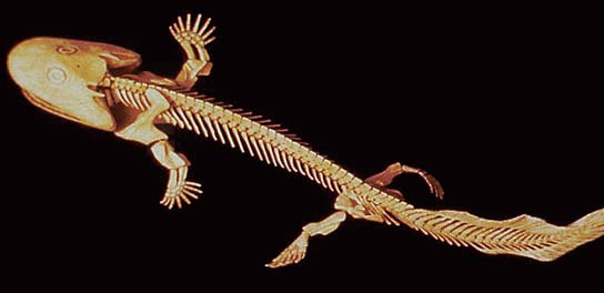
Clack et al. set out to build a method of digital reconstruction that builds upon previous methods, giving greater geometric accuracy. The methods revolve around using a digital mesh obtained through laser or C-T scanning as a model for a landmark-based geometric reconstruction. The sample specimen is a vertebra from the infamous tetrapod fossil Acanthostega. Only one half of the vertebra is actually preserved, therefore this was digitally reconstructed and attached to its mirror image, creating a bilaterally symmetrical three-dimensional element.
Landmark selection involved a mixture of Type 1 and Type 2 landmarks; that is topographically homologous points, mixed with sites of geometric significance, such as local maxima or minima of curvature. These were used as the basis for constructing a surficial grid of contour lines describing the medial and lateral geometry of the neural spine. Videos of the processes involved are actually available online, embedded within the article, a really awesome and useful addition, making the whole methodology more transparent and easier to replicate, should you wish. There’s not really much else to say about the methodology; the processes, such as modelling and surface extrapolation are laid out systematically and reasonably easy to understand for anyone with an understanding of the concepts of geometry and fossils.
The resultant reconstructions are high quality, smooth and geometrically faithful in representing the original vertebra in three dimensions, free of any taphonomic deformation or distortion, and with missing parts accurately reproduced. The groups of models created are validated using Procrustes superimposition and principal components analysis, two standard statistical techniques. The first two principal components do appear to have a low explanatory power however (PC-1 = 24.3%), which may be an issue relating to the complexity in the form of the vertebra. The authors are right to discount the use of the thin-plate spline technique, as this is known to be misleading in that the deformation patterns it produces are homogeneous with respect to the landmark configuration, leading to potentially false morphological variation in areas of no data, something which is largely overlooked.

The advantages of the techniques explored here are in the handling style of the models, and their statistical power and accuracy. Furthermore, anyone can conduct or replicate these methods, providing they have access to an initial CT scan. The potential applications are numerous too: digital models of reconstructed elements can give more accurate parameters for biomechanics where data may have been previously extrapolated in a subjective or qualitative manner; it may yield hitherto unknown data for character construction, which may in turn increase the validity of phylogenetic analysis. The landmark mapping procedure may need refinement in terms of increasing the number of points, such as by using semi-landmarks, which will more accurately reconstruct the surface geometry and open the way for other statistical procedures.
The study represents a great step forward though in accurate specimen reconstruction, and reveals another field in which the power of geometric morphometric techniques is unparalleled. A limitation could be that to reconstruct missing parts, you have to have an idea of what the gross geometry is, meaning at least one half of a bilaterally symmetrical element must be present. This means that if you wanted to reconstruct the neural spine for example, it would be impossible if the whole part was absent, even if the entire centrum was preserved. This is something that could be integrated in future using close relatives of the species that are being reconstructed.
The low score of the principal component axes could well represent the fact that the measurements are independant one from another and express different aspects of the morphology, rather than a general deformative trend.
Great post, as always, Jon. It all kinda reminds me of what Gould said in his 1991 paper on the BG arthropods. Forgive me for quoting pretty liberally my favourite bits,
I would say that, with digital morphometrics, finally we have the technique that Gould was waiting for. A long time coming, though!
I meant BS – Burgess Shale btw. And here’s a citation for the Gould paper, if you wanted it,
Gould, S.J. “The Disparity of the Burgess Shale Arthropod Fauna and the Limits of Cladistic Analysis: Why We Must Strive to Quantify Morphospace.” Paleobiology (1991): 411–423.
Jon,
I appreciate your interest in pterosaurheresies.com and I recognize your potential by the way you’ve handled your own blog and the subjects therein. I’m very impressed and expect great things from you as time goes by.
Dave